X-rays Map Kingfisher Feather Nanostructure, Revealing Blueprint for Iridescence
This article was written by AI based on multiple news sources.Read original source →
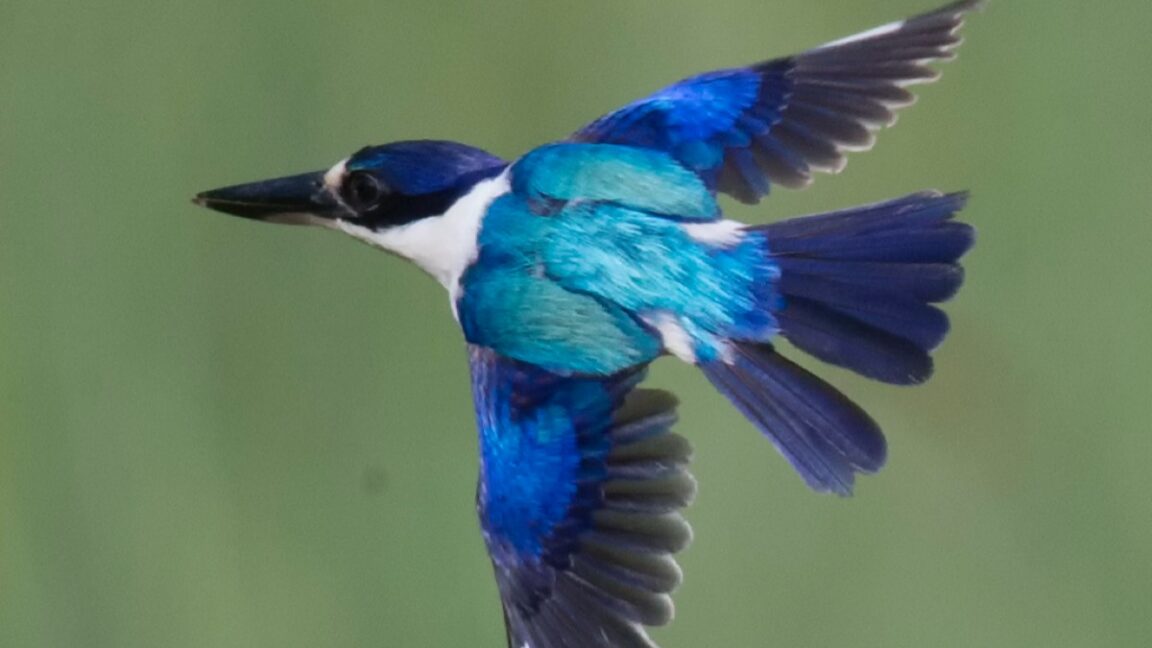
Scientists have unveiled the intricate nanostructure of kingfisher feathers in unprecedented detail, revealing the precise biological architecture behind the bird's dazzling, iridescent plumage. Using powerful synchrotron radiation imaging, a team of researchers has mapped the feather composition at a nanoscale level, discovering a complex, porous, sponge-like framework. This breakthrough analysis provides a definitive look at the physical mechanisms of structural coloration in these birds, moving beyond theoretical models to direct observation. The findings, published in a detailed study, offer a new benchmark for understanding how nature produces vivid colors without pigments, through the manipulation of light itself.
The research centered on applying advanced X-ray imaging techniques typically reserved for materials science to biological specimens. By utilizing synchrotron radiation—intense light generated by particle accelerators—the team could non-destructively peer deep into the feather barbules, the tiny branches that make up a feather's vanes. What they found was not a solid, layered structure often hypothesized, but a three-dimensional network of keratin and air with a highly variable porosity. This nanostructure acts as a photonic crystal, where the spacing and arrangement of the material's tiny gaps interfere with specific wavelengths of light. As light enters this porous matrix, it is scattered and reflected in a way that amplifies certain colors, creating the shimmering blues and greens characteristic of kingfishers. The intensity and hue change with the viewing angle, a hallmark of iridescence, which is directly controlled by the precise geometry of this natural nanostructure.
This detailed structural analysis resolves long-standing questions about the specific biological implementation of structural color in kingfishers. Prior studies often inferred the structure from optical properties or examined limited two-dimensional cross-sections. The synchrotron imaging provides a comprehensive, three-dimensional map, showing how the sponge-like morphology is optimized for light interaction. The variability in pore size and distribution across different parts of the feather likely fine-tunes the optical output, allowing for the complex color patterns seen in the bird's plumage. This represents a significant leap from understanding the principle of structural coloration to documenting its exact physical blueprint in a living organism.
The implications of this discovery extend far beyond ornithology, opening new pathways for biomimetic material science. Engineers and chemists have long sought to replicate structural color to create vibrant, fade-resistant paints, coatings, and textiles without using toxic or unstable pigments. The kingfisher's porous nanostructure offers a sophisticated and efficient biological template for such innovation. By mimicking this sponge-like architecture, researchers could develop new synthetic photonic materials that are lighter, more durable, and capable of dynamic color shifts. Potential applications range from advanced anti-counterfeiting features on currency and passports to more efficient reflective displays and even architectural cladding that changes color with environmental conditions. This research effectively provides a high-resolution design guide drawn from millions of years of natural evolution.
Key Points
- 1Synchrotron imaging revealed a porous nanostructure in kingfisher feathers.
- 2This structure creates bright, iridescent colors through structural coloration.
- 3The research could inspire new bio-inspired synthetic pigments and materials.
This discovery provides a precise biological model for structural color, guiding the development of eco-friendly, fade-resistant synthetic pigments and advanced photonic materials.